We provide the following diagnosis procedures at the Pränatalzentrum an der Wien:
Genetic consultation (human genetics)
As a human geneticist, I can provide you with competent advice at an early stage with regards to the potential risk of passing on a disease as well as in the case of existing (genetic) disorders or abnormalities of your baby.
Down syndrome test on the basis of the mother’s blood – non-invasive prenatal test (NIPT)
(from the 11th week)
Using highly developed laboratory methods, genes of the unborn child (fetal DNA), which are found in the mother’s blood, are examined for the three most common genetic disorders – trisomy 21, 18 and 13. For this purpose, a blood sample is taken from the mother to be sent for analysis at a special laboratory in Germany or America. The result is available after approximately 1 week, the detection rate is 99 percent.
It is recommended to combine the taking of blood samples with an ultrasound scan. In the event of abnormalities or an increased nuchal fold of the baby, invasive diagnostics are preferable to the test.
First trimester ultrasound and nuchal translucency scan
(11th-14th week)
With this ultrasound examination, 60 to 70 percent of structural abnormalities can be detected or ruled out. The thickness of the nuchal fold also determines whether the child has an increased risk for Down syndrome or other chromosomal disorders or syndromes or if there is a heart defect.
Combined test
(11th-14th week)
This test includes a comprehensive ultrasound scan of the fetus and a specific chemical analysis of the mother’s blood. The probability of a chromosomal abnormality is quickly and accurately determined from the combination of results. This result also acts as a decision-making tool for whether a placenta or amniotic fluid test is recommended. In addition, the risk of pre-eclampsia is calculated.
Placenta and amniotic fluid test
(from the 11th or 16th week)
With the examination of fetal cells (of the child), which are sampled by means of puncturing the placenta or the amniotic fluid of the mother, chromosomal disorders of the child can be determined with 100% certainty. Given the relatively high risk of miscarriage (1:100), the two procedures are only used in the event that one or more results of another examination method (e.g. combined test or nuchal translucency scan) indicate an increased risk.
Organ screening
(20th-22nd week)
As part of a detailed, complex ultrasound scan, the child is carefully examined from head to toe. Around 90 percent of all structural abnormalities of bones and organs are identified as a result. The often neglected examination of the heart during conventional ultrasounds is a particular concern of mine.
Cervical ultrasound
(from the 22nd week)
With this vaginal ultrasound scan of the inner cervix, any inclination towards premature birth can be determined.
Well-being ultrasound
(from the 24th week)
This scan serves to determine whether the child is doing alright and is well. The size and circumference of the head and abdomen are also measured. Furthermore, the blood flow of the placenta, umbilical cord as well as other blood vessels of the child are examined and the amount of amniotic fluid checked.
3D ultrasound
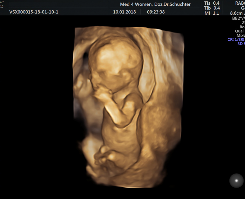
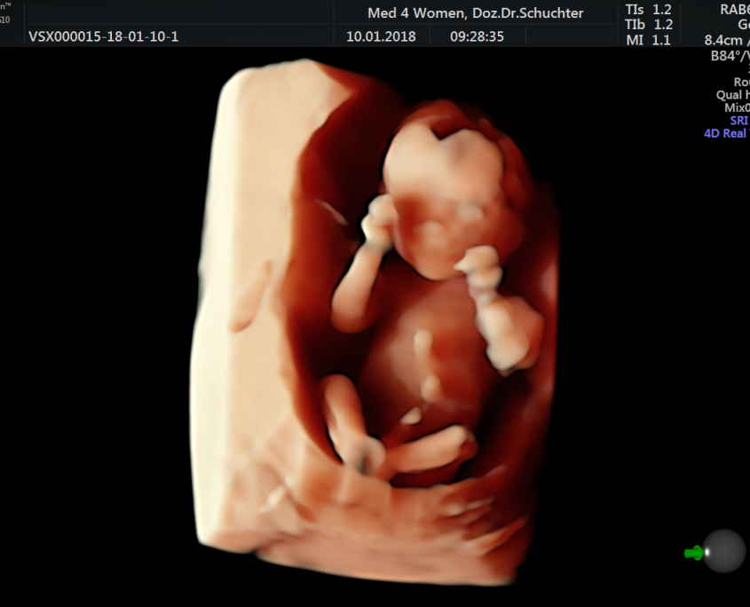
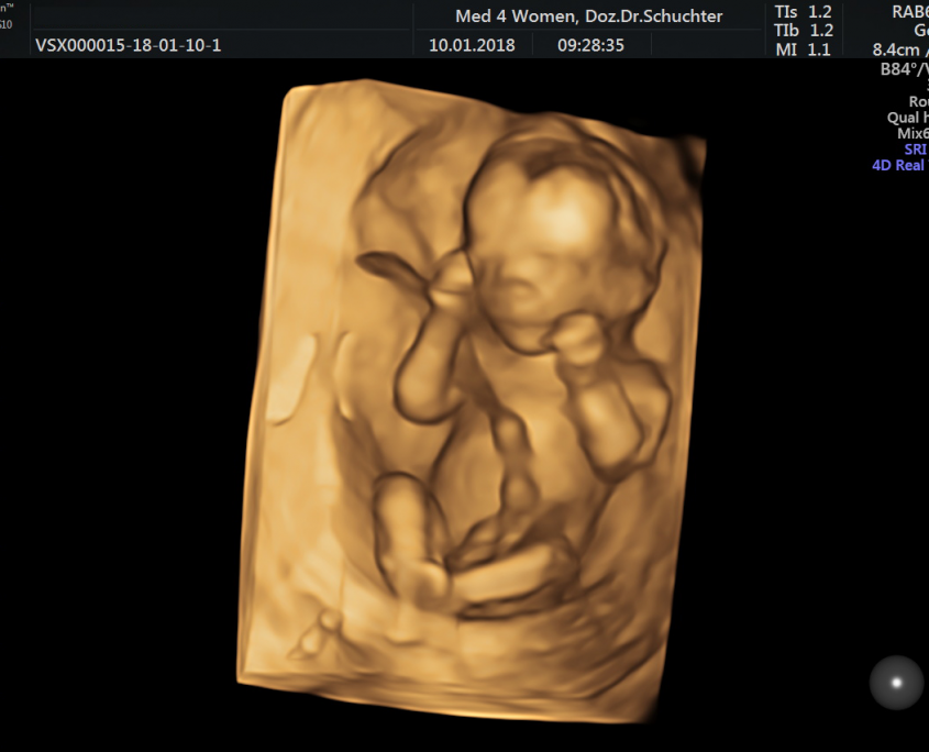
The three-dimensional ultrasound offers expectant parents the opportunity to get a ‘realistic’ picture of their child in the womb. The best gestational age for images is between the 12th and 16th week of pregnancy (for full body images of the baby) and between the 25th and 33rd week (for detailed images).
In principle, a 3D ultrasound functions in the same way as a ‘traditional’, normal ultrasound. The difference is that not only one image but rather many different sectional images are created during the 3D ultrasound and then converted into a three-dimensional image by a computer – then you can see your baby up-close on the screen.
These specific ultrasound examinations are carried out by my colleague Dr. Michaela Wurnig on Wednesdays at med4women in Taubstummengasse following arrangement by telephone on +43 (0)650 731 91 37 (michaela.wurnig@med4women.at). She has many years of experience in the field of 3D ultrasound and was also involved in the creation of the book “Ein Kind entsteht” by Lennart Nilsson (Mosaik publishing house, 2003).
All photos and video sequences taken in the course of the examinations are then immediately given to you on a USB stick.
